Co-author by: Carly Barbon, M.A., SLP, Reg. CASLPO; Ashwini Namasivayam, M.H.Sc., S-LP(C), Reg. CASLPO
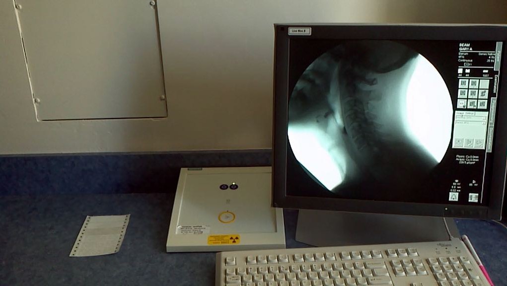
Videofluoroscopy (VFSS) is often referred to as a gold standard assessment method. This method of assessment is ideal for revealing both the anatomy and physiology of swallowing, and for demonstrating pathophysiology in people with dysphagia. VFSS is often used to confirm clinical suspicions with regards to the presence or absence of aspiration, in addition to directly observing physiology and important details that are not visible at the bedside. However, the purpose of the VFSS should not simply be to detect aspiration, but also to explore and confirm the effectiveness of selected approaches to intervention.
Standardization of the stimuli used in videofluoroscopy is critical, as this allows for a common frame of reference across patients, and for comparison between an initial video and one performed after a course of therapy. The contrast agent itself (typically barium), how it is prepared, the order of stimulus presentation, and the volumes to be tested are all important aspects of the examination to consider. One must also be careful regarding technical aspects regarding fluoroscopy and image registration. Best practice recommendations are to generate the maximum number of images possible per second to ensure that brief events are not missed. In North America, optimum frame rate is defined either ascontinuous fluoroscopy or pulsed fluoroscopy at 30 pulses per second, with registration on a recording system at 30 frames per second.
Barium is the most commonly used contrast agent for VFSS in North America. Varibar®, a product line available only in the United States, is made specifically for oropharyngeal swallow examinations. It comes in different consistencies, all containing the same concentration of barium (40% weight/volume). For clinicians who cannot access Varibar®, our lab is currently developing standard recipes for VFSS using EZ-Paque® barium powder. Once recipes have been tested, they will be shared at www.steeleswallowinglab.ca.
It is imperative to understand the properties of the barium used because higher concentration barium is intended to coat the walls of the gastrointestinal system (Steele et al., 2013). If a healthy individual swallows Liquid Polibar®, which is 100% weight/volume concentration, coating should be expected along the entire oropharynx. It is important not to confuse this coating for residue. Lower concentrations of barium like Liquid EZ-Paque® or Varibar® leave less coating behind. Barium diluted with water to a 20% weight/volume concentration is believed to be as close to a true thin liquid as possible, while remaining radiopaque but leaving minimal coating on the walls of the oropharynx.

In addition to standardized stimuli, a videofluoroscopy protocol is also important. The MBSImp (Martin-Harris et al., 2008) is an example of one such approach, requiring extensive training, and including the analysis of 17 different physiological aspects of a swallow. At the Toronto Rehabilitation Institute, a hospital within the University Health Network in Toronto, Canada, a slightly different standard protocol is in place. All questions are answered in a maximum of 16 boluses to avoid excess radiation. A saliva swallow is often conducted first to allow for evaluation of structural movement. Second, the patient is asked to hold a 10 ml volume of thin liquid in their mouth for 5 seconds to challenge oral bolus control. Six core teaspoon swallows are then included, three of thin and three of spoon-thick barium. The use of three repeated boluses of each stimulus allows us to capture representative swallowing function in patients. The remainder of the protocol allows the clinician to explore different consistencies, volumes, or techniques in efforts to improve swallowing function.
There are several different features that can be rated in videofluoroscopy. These are best separated into functional and physiological parameters, which help to understand breakdowns in function. The first concern is swallow safety (penetration and/or aspiration). The second is swallow efficiency, which can be measured using scales of residue severity. These two measures are the most critical for clinicians in regular practice. Other physiological parameters can also be considered to aid in the understanding of dysfunction.
The 8-point Penetration Aspiration Scale (PAS) has become the standard tool for describing the severity of aspiration (Rosenbek, 1996). The scale is broken down into eight different levels but is frequently reduced to a binary scale, where any score higher than 2 is considered abnormal. This reflects the fact that whenever material enters the supraglottic space and stays there, it is a risk for eventual aspiration.
Judging the severity of residue is challenging, and until recently, most clinicians and researchers have used subjective ordinal scales that capture how full a space appears to be. A new scale for measuring residue severity called the Normalized Residue Ratio Scale (NRRS) has been introduced by Pearson and colleagues (2013). Using ImageJ, a free analysis software, the area of residue is traced on an image and divided by the area of the space it is filling. The NRRS also incorporates an anatomical scalar reference to account for individual differences in size.

Timing is also critical when analyzing a VFSS. To determine underlying physiological parameters that explain dysphagia, the clinician must have clear definitions for each of the events of interest. For example, by subtracting the time of bolus entry into the pharynx from the time of airway closure, one can calculate the length of the time that the bolus is sitting in the pharynx while the airway is open, which may explain one important mechanism of aspiration.
Specific measurements on frames of interest can be made to evaluate swallowing kinematics. For example, we typically identify the frame of peak hyoid position, and measure the position of the hyoid relative to a fixed point on the cervical spine. Recent evidence suggests that kinematic measures need to be adjusted for differences in participant height using a cervical spine scalar.
Finally, it is important to link the kinds of problems seen in videofluoroscopy to treatment planning. When following a standardized protocol for testing and analysis, it is possible to begin developing a matrix, where certain approaches to management are indicated by certain profiles of pathophysiology. We recommend this approach to guide the selection of techniques that are most-suited to the specific nature of a person’s dysphagia, when probing treatment effectiveness in VFSS.
For more information on the exciting research from Dr. Steele’s Lab visit: www.steeleswallowinglab.ca.
About the Authors
Dr. Catriona M. Steele, Ph.D., CCC-SLP, BCS-S, ASHA Fellow is a Professor in the Department of Speech-Language Pathology at the University of Toronto and Director of the Swallowing Rehabilitation Research Laboratory at the Toronto Rehabilitation Institute – University Health Network (www.SteeleSwallowingLab.ca).
Co-authors Ashwini Namasivayam and Carly Barbon are both Ph.D. students in Dr. Steele’s lab.
References
- Martin-Harris, B., Brodsky, M.B., Michel, Y., Castell, D.O., Schleicher, M., Sandidge, J., Maxwell, R., & Blair, J. (2008). MBS measurement tool for swallow impairment – MBSImp: establishing a standard. Dyphagia, 23(4), 392-405.
- Pearson, W.G., Molfenter, S.M., Smith, Z.M., & Steele, C.M. (2013). Image-based measurement of post-swallow residue: the normalized residue ratio scale. Dysphagia, 28(2), 167-177.
- Rosenbek, J. C., Robbins, J. A., Roecker, E. B., Coyle, J. L., & Wood, J. L. (1996). A penetration-aspiration scale. Dysphagia, 11, 93-98.
- Steele, C. M., Molfenter, S. M., Peladeau-Pigeon, M. & Stokely, S. L. (2013). Challenges in preparing contrast media for videofluoroscopy. Dysphagia, 28(3), 464-467.


