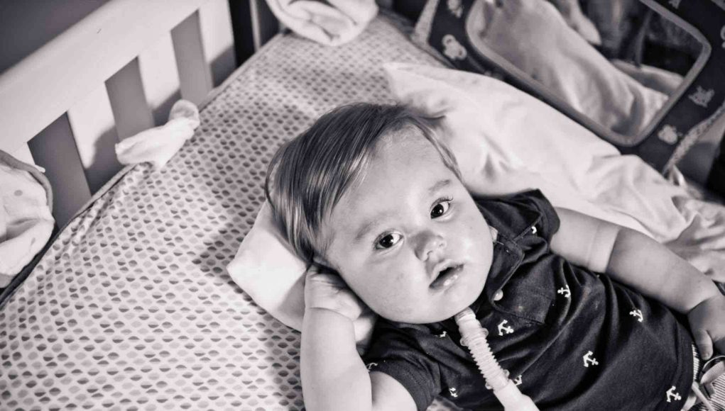
Dysphagia in children and young adults with neuromuscular disorders (NMD) is an important clinical issue, resulting in malnutrition, dehydration and/or aspiration pneumonias. It may prohibit social participation and cause fear of choking. Therefore, assessment and treatment recommendations are important to improve safe and efficient feeding management.
In children the most obvious sign of a neuromuscular disorder is hypotonia due to muscle weakness. Neuromuscular disorders are broadly subdivided in myopathies and neuropathies. The most common neuromuscular disorders in children are Duchenne muscular dystrophy (DMD) and spinal muscular atrophy (SMA), followed by hereditary motor sensory neuropathy and myotonic dystrophy (table 1) [1, 2].
Feeding disorders and dysphagia in children with neurodevelopmental disabilities vary widely. Children with cerebral palsy (CP) are at risk of aspiration of thin fluid [3, 4]. Recommendations for safe oral feeding in this patient group are traditionally focused on changing posture during feeding and thickening fluids. However, different patterns in swallowing problems were found in children with CP and children with neuromuscular disorders (NMD) [5]. In children with NMD malnutrition [6], impaired chewing performance and oral phase problems [7, 8], choking and post-swallow residue [8-10] are described. The oral motor problems are often complicated by structural impairments [11].
In the literature a wide range of swallowing problems in children with NMD is reported, related to different factors: (1) feeding problems in the neonatal period differ from childhood, because of different food substances or the use of utensils; (2) dysphagia can be observed in the oral and/or pharyngeal phase; (3) the stability or progressiveness of the disease differ from one disease to another; (4) bulbar involvement of oral or pharyngeal muscles or a combination varies among different NMD’s in childhood. To start appropriate rehabilitation in this patient group, mechanism based descriptions of the specific dysphagia are needed [12].
For example the dysphagia in children with SMA type II, described in our study [9], was assessed with surface EMG of the submental muscle group and a videofluoroscopic swallow study (VFSS) in two different postures. The dysphagia in SMA II is due to neurologic dysfunction of lower motor neurons of the cranial nerves from the brainstem, affecting both the force and the efficacy of the tongue and submental muscle group, in combination with a compensatory retracted head posture. These findings have led to an integrated treatment advice to adopt an adjusted posture during meals and to drink water after meals to prevent aspiration pneumonias.
The dysphagia in boys and (young) adults with DMD was assessed with sEMG of the submental muscle group and anterior tongue pressure during swallowing, quantitative muscle ultrasound (QMUS) and a VFSS, in 24 DMD patients, aged 5 – 40 years [10]. The participants were divided in three groups, based on the stage of their disease (9): early and late ambulatory stage (AS, N=6), early non-ambulatory stage (ENAS, N=7), and late non-ambulatory stage (LNAS, N=11).
Several studies have shown that changes in muscles, caused by neuromuscular disorders, can be detected by muscle ultrasound. With quantitative muscle ultrasound (QMUS) echo intensity and muscle thickness can be described and compared to normal values [13]. A study of our group revealed that QMUS was also feasible for oral muscles, i.e. m. digastricus, m. geniohyoid, and tongue muscles [14] (Figure 1). Echo intensity and thickness were described as Z-scores and used to quantify healthy and affected muscles.
With QMUS it was shown that the echo intensity of the oral muscles in DMD gradually increased, starting in the AS group with the geniohyoid muscle. This increased echo intensity reflects the structural changes in muscles caused by DMD, related to muscle weakness [15]. The echo intensity of the oral muscles showed the same gradual involvement as observed in various skeletal muscles, but at a later onset. The first dystrophic change of the geniohyoid muscle was found in the AS, whereas dystrophic changes of the digastric muscles and the superior longitudinal tongue muscle were only seen in the LNAS. On the VFSS none of the patients showed direct aspiration. Nevertheless, oral phase problems and pharyngeal post-swallow residue were often observed, mostly in the LNAS and most frequently with thick liquid and solid food. These findings indicate that the complex function of swallowing, with involvement of the oral and pharyngeal muscles, is gradually affected over time. As a consequence, feeding and swallowing problems develop slowly, which requires dedicated professional attention already in the early stages of DMD.
Patients with SMA II and DMD rarely spontaneously complain about swallowing problems. Both diseases are slowly progressive and, as a consequence, patients may easily adapt their feeding and swallowing habits to their reduced oral strength without realizing this gradual change. This phenomenon has also been reported in adults with a progressive neuromuscular disorder [16]. It will lead to compensations (either adequate or inadequate) with possible positive or adverse side effects [17]. Because patients do not easily report their complaints, it hampers the possibility to benefit from a feeding and swallowing assessment and subsequent recommendations. It emphasizes the importance to pay attention to changing feeding and swallowing habits that may mask dysphagia already in the early stages of neuromuscular disorders.
In general, rehabilitation of patients with neuromuscular disorders is focused on both behavioral compensation and task-specific restorative training, taking into account the underlying pathophysiology. The primary aim in the rehabilitation of feeding and swallowing problems is to achieve safety, i.e. the prevention of choking and aspiration pneumonia. Essential differences between children with central and peripheral neurologic disorders need to be taken into account and call for intensive collaboration between the pediatric neurologist, rehabilitation physician and speech-language therapist. Choking in children with CP has a different pathophysiological origin than in children with a neuromuscular disorder and must, therefore, be treated differently. In children with CP, thickened liquids will prevent aspiration during swallowing, whereas in children with neuromuscular disorders thickened liquids are not recommended, because they aggravate post-swallow residue. Cleaning the oropharyngeal area with water after meals is important.
For SLTs working with children and young adults with a neuromuscular disorder, it is important to carefully consider the underlying mechanisms of oral and pharyngeal dysphagia to be able to provide mechanism-based recommendations for efficient and safe swallowing.
References
- Dubowitz, V., Muscle Disorders in Childhood
. 2000, London: W.B. Saunders Company LTD.
- Yang, M.L. and R.S. Finkel, Overview of paediatric neuromuscular disorders and related pulmonary issues: diagnostic and therapeutic considerations. Paediatr Respir Rev, 2010. 11(1): p. 9-17.
- Arvedson, J., et al., Silent aspiration prominent in children with dysphagia. Int.J.Pediatr.Otorhinolaryngol., 1994. 28(2-3): p. 173-181.
- Arvedson, J.C., Assessment of pediatric dysphagia and feeding disorders: clinical and instrumental approaches. Dev.Disabil.Res.Rev., 2008. 14(2): p. 118-127.
- van den Engel-Hoek, L., et al., Children With Central and Peripheral Neurologic Disorders Have Distinguishable Patterns of Dysphagia on Videofluoroscopic Swallow Study. J Child Neurol, 2013.
- Tilton, A.H., M.D. Miller, and V. Khoshoo, Nutrition and swallowing in pediatric neuromuscular patients. Semin.Pediatr.Neurol., 1998. 5(2): p. 106-115.
- Aloysius, A., et al., Swallowing difficulties in Duchenne muscular dystrophy: indications for feeding assessment and outcome of videofluroscopic swallow studies. Eur.J.Paediatr.Neurol., 2008. 12(3): p. 239-245.
- Pane, M., et al., Feeding problems and weight gain in Duchenne muscular dystrophy. Eur.J.Paediatr.Neurol., 2006. 10(5-6): p. 231-236.
- Engel-Hoek, L.v.d., et al., Dysphagia in spinal muscular atrophy type II: more than a bulbar problem? Neurology, 2009. 73(21): p. 1787-1791.
- van den Engel-Hoek, L., et al., Oral muscles are progressively affected in Duchenne muscular dystrophy: implications for dysphagia treatment. J Neurol, 2013. 260(5): p. 1295-303.
- Bagnall, A.K., M.A. Al-Muhaizea, and A.Y. Manzur, Feeding and speech difficulties in typical congenital Nemaline myopathy, in Advances in Speech-Language Pathology2006. p. 7-16.
- van der Wilt, G.J. and G.A. Zielhuis, Merging evidence-based and mechanism-based medicine. Lancet, 2008. 372(9638): p. 519-520.
- Pillen, S., et al., Quantitative skeletal muscle ultrasound: diagnostic value in childhood neuromuscular disease. Neuromuscul.Disord., 2007. 17(7): p. 509-516.
- van den Engel-Hoek, L., et al., Quantitative ultrasound of the tongue and submental muscles in children and young adults. Muscle Nerve, 2012. 46(1): p. 31-37.
- Jansen, M., et al., Quantitative muscle ultrasound is a promising longitudinal follow-up tool in Duchenne Muscular Dystrophy. Neuromuscul.Disord., 2012. 22(4): p. 306-317.
- Swart, B.d., Speech therapy in patients with neuromuscular disorders and Parkinson’s disease., 2006, Radboud University Nijmegen, the Netherlands.
- de Swart, B.J., et al., Ptosis aggravates dysphagia in oculopharyngeal muscular dystrophy. J.Neurol.Neurosurg.Psychiatry, 2006. 77(2): p. 266-268.
Links of Interest
- PhD Medical SciencesLink to Thesis: http://repository.ubn.ru.nl/bitstream/handle/2066/107646/107646.pdf?sequence=1
- Link to hospital webpage (Dutch only):
radboudumc.nl/ZORG/AFDELINGEN/REVALIDATIE/LOGOPEDIE/Pages/Logopediekinderen.aspx


