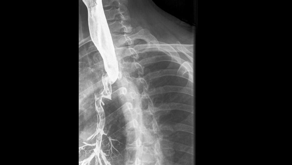
Feature Image Credit: Case courtesy of Dr Hidayatullah Hamidi, Radiopaedia.org. From the case rID: 51356
Introduction
Modified barium swallow studies (MBSS) typically involve use of a small palette of barium sulfate-based preparations of different textures/viscosities. Clinicians who prioritize inter-patient and inter-institution consistency of testing and results may use a suite of preparations of standardized viscosities designed for the MBSS, such as Varibar® (Bracco Diagnostics Inc., Monroe NJ).1 Others may use one or more homemade preparations, mixing other BaSO4 formulations with various foods or food additives. Ideally, the MBSS would be conducted using a clinically validated protocol, such as the Modified Barium Swallow Impairment Profile (MBSImP®).2,3
But what if the patient has a potentially serious complicating issue? What if there is a perforation of the esophagus? What if there is a high risk of aspiration, or of serious consequences from aspiration? What contrast media can or should one use? To answer such questions, one must understand:
- The features of the potentially serious complicating issues;
- What the available contrast media truly are – not just their names, but their natures;
- The effects of the various contrast media if exposed to the body tissues or airways.
Then, one must balance an accurate understanding of the risks vs. benefits of an MBSS to arrive at an informed decision on how to proceed.
Perforations, Penetrations and Fistulas
Broadly, a perforation is an “abnormal opening in a hollow organ or viscus.”4 However, when considering GI tract ulcerations, a further distinction is made: ulcers are penetrating if they erode through the bowel wall without free perforation and leakage into a body cavity,5 and perforating if they do erode into a body cavity. The majority of the esophagus is located in the mediastinum,6 and does not pass through a body cavity, so an ulceration or tear would result in penetration of swallowed material into interstitial tissues.
However, an abnormal opening in a hollow organ or viscus can connect directly into another hollow organ or viscus, forming a fistula. GI tract fistulas can arise from surgery, inflammation, cancer, or penetrating, tearing, caustic or radiation injury.7 The adjacent hollow organ to which an esophageal fistula may connect is the trachea. Acquired tracheoesophageal (T-E) fistulas most commonly occur at the junction of the cervical and thoracic esophagus; about half are cancerous in origin.8 Congenital T-E fistulas are usually malformations associated with esophageal atresia in newborns; however, the rare H-type fistula may present in adults, in whom it can cause severe morbidity and mortality.9
Contrast Media for Radiographic Evaluation of the Esophagus
Two broad categories of contrast media are available for use in radiographic evaluation of the esophagus: (1) barium sulfate suspensions, and (2) iodinated contrast media solutions.
Barium sulfate (BaSO4) is a chemically inert, insoluble mineral. It can only be liquified as a suspension, with the help of emulsifiers, stabilizers, dispersants, laxatives, sweeteners, thickeners and preservatives. (Plant gum [e.g., acacia, tragacanth] is added when mucosal coating is desired.10) It is these “excipients” – mostly common food additives – that give BaSO4 preparations most of their variable chemical properties; the BaSO4 moleculeonly provides physical density [g/mL] and radiographic opacification (a.k.a. “radiodensity”).
A BaSO4 product is formulated for a specific purpose – and may be unsuitable when used for something else. Mucosal coating is necessary for double-contrast imaging11 – but is very problematic in the MBSS, where it may give a false appearance of pharyngeal residue. High radiodensity allows visualization of a thin layer of mucosal coating11 – but provides too much opacification for single-contrast studies, including the MBSS. A suite of products with specific textures/viscosities may be formulated for the MBSS,1,2,3 but not to find a peptic ulcer.
Iodinated contrast media (ICM) are aqueous solutions, in which the contrast molecules are fully dissolved in water, typically along with small amounts of a few additives.12,13 ICM are typically categorized as either high-osmolar (HOCM) or low-osmolar (LOCM). HOCM were developed first. Their iodine-containing molecules are ionic, with a net negative electrical charge in solution, requiring the presence of positively-charged companion molecules, usually sodium ions (Na+) and/or meglumine.12,13 This profusion of particles leads to a high osmolality at radiographically useful concentrations. Therefore, HOCM will osmotically draw water from the body into the bowel lumen – and, if aspirated, into the lungs, which is harmful.13 Conversely, LOCM (with one exception) are nonionic, thus having no associated balancing molecules in solution. While their osmolalities are still higher than blood’s, they are low enough to usually avoid significant physiologic adverse effects.14,15
Contrast medium choice – What to Use and When
Perforation. All BaSO4 preparations, including the Varibar® products,1 are contraindicated for use when perforation is known or suspected. Insoluble and chemically inert, BaSO4 particles are neither toxic nor allergenic, but they do stimulate an inflammatory reaction in body tissues. In 1977, Goldner and Adams used light and electron microscopy to study the response of certain circulating white blood cells to BaSO4 particles.16 They saw them mature into tissue macrophages that engulfed the particles and organized into dense granulomas, which are inflammatory masses that form when the immune system attempts to wall off foreign matter it cannot eliminate.
The results of an animal model of esophageal perforation published by Vessal et al17 were summarized by Selzer et al18 in a comprehensive 1979 review. Various mixtures of human mouth flora, a BaSO4 preparation and Gastrografin® (Bracco Diagnostics Inc., Monroe NJ) were instilled into cat mediastina via the esophagus. Small amounts of mouth flora produced inflammation, and larger amounts were commonly lethal. The BaSO4 preparation induced granuloma formation, but when mixed with mouth flora had no added deleterious effect over the flora alone. Gastrografin produced no reaction.
The ACR Manual on Contrast Media 202014 summarizes current thinking on the issue:
“The most serious complication from the use of barium in the GI tract is leakage into the mediastinum or peritoneal cavity [1].19 The potential complications of a barium leak depend on the site from which the spill occurs. Esophageal leakage may cause mediastinitis. Stomach, duodenal, and small intestinal leakage may result in peritonitis. Escape of barium from the colon, where the bacterial count is highest, carries high mortality (with the mortality likely primarily related to leakage of stool).”(Italics added.)
This complication is most simply avoided by using water-soluble ICM: 14
“Water-soluble contrast media are absorbed rapidly from the interstitial spaces and peritoneal cavity, a feature that makes them uniquely useful in examining patients with a suspected perforation of a hollow viscus. No permanent deleterious effects from the presence of water-soluble contrast media in the mediastinum, pleural cavity, or peritoneal cavity have been shown to occur [14].”17
In considering the MBSS, the use of only water-soluble ICM is quite limiting, because these agents are all thin liquids. To produce any other texture/viscosity, one must add foods or food additives, which themselves would be contraindicated with a perforation. In sum, the GI perforation should be addressed before a full swallowing evaluation is attempted.
Fistulas. Contrast media passing through fistulas typically do not suffuse into the surrounding interstitial tissues, but follow the path of least resistance connecting the hollow structures. So, there may be little or no adverse effect from use of a BaSO4 preparation for diagnosis of an enteric fistula; in one trial, 625 patients with Crohn’s disease (CD) were assessed for internal fistulas using barium enema and enteroclysis, without undue occurrence of adverse events.20 Fistulas may also connect the GI tract to the urinary bladder or other hollow viscera, and BaSO4 examination even of these may be relatively uneventful; one review of 500 CD patients found that visualization of an enterovesical (bowel to bladder) fistula on barium enema was not associated with adverse outcome.21 However, since esophageal fistulas will most likely connect to the trachea, aspiration is the principal consideration in evaluating them.
Aspiration. Aspiration and its potential serious consequences (e.g., choking, pulmonary edema, pneumonia) is not a consideration unique to BaSO4 preparations. Aspirated food, drink or medication may physically block the airways, introduce harmful mouth flora, be caustic or hypertonic, and/or have viscous, fibrous or particulate matter that is pro-inflammatory and difficult to clear from the lungs. In their study of the economic burden of dysphagia, Patel et al noted that “consequences of untreated and unrecognized dysphagia can be profound, including malnutrition, volume depletion, quality of life issues, and aspiration, which may ultimately be the pivotal factor that precipitates a decline in a patient’s outcome.”22 (Italics added.)
Aspiration of BaSO4 was extensively studied and reported decades ago.15,17 It was found that introduction of smaller amounts of uncontaminated BaSO4 into the bronchial tree is likely to be clinically harmless if it does not obstruct major bronchi or contain regurgitated stomach contents; coughing and ciliary action usually removes most of the material quickly (unless the bronchial lining is damaged14,23). However, aspiration of large volumes can cause major bronchial obstruction, or lead to alveolar deposition with inflammatory granuloma formation, while aspirated gastric contents induce chemical bronchitis and pneumonia.15
But in patients in whom there is a concern of large volume aspiration, the use of iodinated HOCM indicated for oral administration, such as Gastrografin12 or MD-Gastroview® (Guerbet LLC, Princeton NJ)13 is certainly not the answer. As the ACR Manual states:14
“HOCM may, if aspirated, cause life-threatening pulmonary edema [2,17,18]. Therefore, if water-soluble contrast media are to be used in patients at risk for aspiration, low-osmolality or iso-osmolality contrast media are preferred, as these contrast agents, if aspirated, are associated with only minimal morbidity and mortality [17].” 15,18,24
But all LOCM are thin liquids. That works for a single-contrast esophagram, or for studying the ability to swallow thin liquid – but what about the full MBSS? Wasn’t the patient referred to you for a thorough swallowing evaluation because of oropharyngeal dysphagia, which causes aspiration? Knowledgeably balancing the potential risk of complications from the aspiration of BaSO4 preparations (which is low with controlled, low-volume aliquots of an uncontaminated preparation), with the benefits of properly evaluating a serious condition that leads to poor outcomes, will help guide you out of this dilemma, toward optimal patient care.
References
- Full prescribing information for Varibar® Thin Liquid (Barium Sulfate) for Oral Suspension, 81% w/w, Varibar® Nectar (Barium Sulfate) Oral Suspension, 40% w/v, Varibar® Thin Honey (Barium Sulfate) Oral Suspension, 40% w/v, Varibar® Honey (Barium Sulfate) Oral Suspension, 40% w/v and Varibar® Pudding (Barium Sulfate) Oral Paste. Bracco Diagnostics Inc., Monroe NJ.
- Martin-Harris B, Brodsky MB, Michel Y, Castell DO, Schleicher, Sandidge J, Maxwell R, Blair J. MBS Measurement Tool for Swallow Impairment – MBSImp: Establishing a Standard. Dysphagia. 2008; 23: 392-405.
- Martin-Harris B, Humphries K, Garand KL. The Modified Barium Swallow Impairment Profile (MBSImP©) – Innovation, Dissemination and Implementation. Perspectives of the ASHA Special Interest Groups SIG 13. 2017; 2(4): 129-138.
- Medical Dictionary for the Health Professions and Nursing, © Farlex 2012. Accessed at
https://medical-dictionary.thefreedictionary.com/perforation. - Pasumarthy L, Kumar RR, Srour J, Ahlbrandt D. Penetration of Gastric Ulcer into the Splenic Artery: A Rare Complication. Gastroenterology Res. 2009 Dec; 2(6): 350–352.
- Chaudry SR, Bordoni B. Anatomy, Thorax, Esophagus. StatPearls [on-line]. Accessed at https://www.ncbi.nlm.nih.gov/books/NBK482513/.
- Gastrointestinal fistula. MedlinePlus (on-line). U.S. National Library of Medicine. Accessed at https://medlineplus.gov/ency/article/001129.htm.
- Diddee R, Shaw IH. Acquired tracheo-oesophageal fistula in adults. Continuing Education in Anaesthesia Critical Care and Pain. 2006 Jun; 6(3): 105-108.
- Suen HC. Congenital H-type tracheoesophageal fistula in adults. J Thorac Dis. 2018 Jun; 10 (Suppl 16): S1905-S1910.
- Data on file. Bracco Diagnostics Inc., Monroe NJ.
- Pickhardt PJ, Kim DH. CT Colonography: Principles and Practice of Virtual Colonoscopy. ©2009 Elsevier Inc. Accessed at: https://www.sciencedirect.com/topics/medicine-and-dentistry/double-contrast-barium-enema
- Gastrografin® full prescribing information. Bracco Diagnostics Inc., Monroe NJ
- MD-Gastroview® full prescribing information. Guerbet LLC, Princeton NJ
- ACR Committee on Drugs and Contrast Media. ACR Manual on Contrast Media 2020.
©2020 American College of Radiology - Gelfand DW. Complications of gastrointestinal radiologic procedures: I. Complications of routine fluoroscopic studies. Gastrointest Radiol. 1980; 5: 293-315.
- Goldner RD, Adams DO. The structure of mononuclear phagocytes differentiating in vivo. III. The effect of particulate foreign substances. Am J Pathol. 1977 Nov; 89(2): 335-350.
- Vessal K, Montali RJ, Larson S, Chaffee V, James E. Evaluation of barium and Gastrografin as contrast media for the diagnosis of esophageal ruptures or perforations. Am J Roentgenol Radium Ther Nucl Med. 1975 Feb; 123(2): 307-319.
- Selzer SE, Jones B, McLaughlin GC. Proper choice of contrast agents in emergency gastrointestinal radiology. CRC Critical Rev Diagn Imag. 1979 Nov; 12(1): 79-99.
- Ott DJ, Gelfand DW. Gastrointestinal contrast agents. Indications, uses, and risks. JAMA 1983; 249: 2380-2384.
- Maconi G, Sampietro GM, Parente F, Pompili G, Russo A, Cristaldi M, Arborio G, Ardizzone S, Matacena G, Taschieri AM, Porro GB. Contrast radiology, computed tomography and ultrasonography in detecting internal fistulas and intra-abdominal abscesses in Crohn’s disease: a prospective comparison study. Am J Gastroenterol. 2003 Jul; 98(7): 1545-1555.
- Margolin ML, Korelitz BI. Management of bladder fistulas in Crohn’s disease. J Clin Gastroenterol. 1989 Aug; 11(4): 399-402.
- Patel DA, Krishnaswami S, Steger E, Conover E, Vaezi MF, Ciucci MR, Francis DO. Economic and survival burden of dysphagia among inpatients in the United States. Dis Esophagus. 2018 Jan 1; 31(1): 1-7.
- Gelfand DW, Ott DJ. Barium sulfate suspensions: an evaluation of available products. AJR 1982: 138: 935-941.
- Reich SB. Production of pulmonary edema by aspiration of water-soluble nonabsorbable contrast media. Radiology. 1969; 92: 367-370.


